|
Invasive Aspergillus
Reliable techniques to confirm a diagnosis in patients with pulmonary or disseminated infection are currently not well established and impede successful management. Biopsy of skin lesions can reveal fungal hyphae, although the organism may not grow in culture media. Cultures of sputum or bronchoalveolar lavage (BAL) fluid are fairly specific (>80% in neutropenic patients); unfortunately, the sensitivity in proven cases is only 25% to 50% [51]. A negative culture from respiratory secretions has little meaning, as often the infection has to be well advanced before a positive culture is obtained. However, a positive culture in an immunocompromised patient suggests that the patient may already be infected or at high risk of becoming infected.
Antibody detection is not useful in most patients, as poor immune function and the speed of onset of the infection mean that antibody titers are low. Serologic methods to detect circulating free antigens or immune complexes have been or are under development using ELISA or radioimmunoassay [52]. Most techniques focus on detecting galactomannan, a component of the fungal cell wall. Galactomannan-based serologic diagnostics have now been intensively studied in vivo and in humans and appear very powerful [53, 54]. This marker can be used for both diagnosing and monitoring response. The current draft of the EORTC/MSG diagnostic criteria [55] includes a positive Aspergillus antigen as one component that can contribute to the diagnosis of probable aspergillosis. A recent study has shown greater than 90% specificity and sensitivity of a galactomannan assay in prolongedneutropenic and/or steroid-treated patients with hematologic disorders [56]. A latex agglutination method to detect Aspergillus antigen is commercially available in Europe (Pastorex, Sano. Diagnostics Pasteur, France), although its sensitivity and specificity are limited. More recently, a sandwich ELISA using monoclonal galactomannan antibody has been evaluated in high-risk patients, although it is not approved for clinical use in the US (Pastorex, Sano. Diagnostics Pasteur, France). The newest development is the use of polymerase chain reaction (PCR)-based techniques to detect Aspergillus DNA in respiratory secretions and serum, but these techniques have not yet achieved the diagnostic consistency or broad acceptance seen with the galactomannan-based approaches [57]. The likelihood of false-positive results may preclude this tool as a stand-alone method to diagnose fungal infection.
Imaging the lungs of patients in whom Aspergillus infection is suspected can be very useful. Chest radiographs may show suggestive rounded parenchymal densities or pleural-based processes. Air crescents due to cavitation are strongly suggestive [58], but not diagnostic and not usually seen until recovery from neutropenia. High-resolution CT scans provide more information than chest radiography [43]. High-resolution CT shows that the parenchymal densities are angiocentric and also permits appreciation of the so-called halo sign (a zone of low attenuation surrounding these pulmonary nodules) that suggests vascular compromise of the region around the primary process [59]. While not absolutely diagnostic, the finding is certainly very suggestive [43]. More important, a negative high-resolution CT largely excludes the possibility of pulmonary aspergillosis [60].
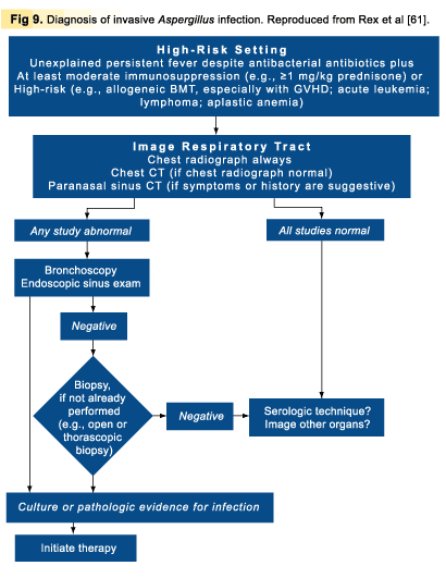
Taken together, these ideas suggest a diagnostic approach based on clinical, microbiologic, and radiologic features (Fig 9) [61].
Invasive Candidiasis
There is a significant lag in developing detection methods for invasive candidiasis largely because there is no convincing gold-standard diagnostic strategy in any form. The spectrum of invasive candidiasis is quite broad, and no single approach has yet emerged.
Current strategies revolve around clinical approaches to detection of disease. The detection of Candida in the bloodstream is one of the simplest and most convincing bits of data, but this is seen in no more than about half of patients in whom invasive candidiasis is strongly suspected [62]. Positive cultures from other sites may represent colonization rather than invasive disease. As a consequence, significant effort has gone into nonculture-based diagnostic strategies. These have included detection of Candida enolase and antibodies to enolase, Candida mannoproteins, beta-glucan, other less defined antigens [63], the candidal metabolic product D-arabinitol [64], and candidal DNA by PCR [50]. All of these methods have shown promise, but none have achieved the degree of diagnostic accuracy that seems needed to justify widespread use. The principal problem is that the positive signal detected by each is strongly present only when the disease is fairly far advanced. Coupled with the rapid pace of invasive candidiasis and the time delay inherent in obtaining test results, these problems have thus far made these tests less than satisfactory outside of highly focused research settings.
Course Number: V035B.043001
This CME Expires on July 1, 2003; no tests will be accepted after this date.
This course is accredited by
The University of Pittsburgh School of Medicine, Center for Continuing Education
|
|